The human body is arguably the most wonderful, complex organisms on this planet. The human body is a single structure but it is made up of billions of smaller structures of four major kinds: cells, tissues, organs and systems.
The body is composed of ten major systems: Skeletal, Muscular, Nervous, Cardiovascular, Lymphatic, Respiratory, Digestive, Endocrine, Urinary, and Reproductive.
The human body consists of three major areas: the head, the trunk and the extremities (limbs). Common names of well known parts of the human body, from top to bottom, are as follows:
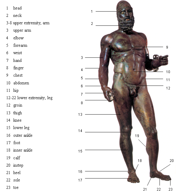
Figure 7: Human body, anterior view
HEAD: FOREHEAD, EYE, EAR, NOSE, MOUTH, TONGUE, TEETH, JAW, FACE, CHEEK ,CHIN, NECK
The front of the head, where the eyes, ears, nose and mouth are located, is called the face. The area above the eyes is called the forehead and below the mouth is the chin.
UPPER LIMB: SHOULDER, ARM, UPPER ARM, ELBOW, FOREARM, WRIST, HAND, FINGERS,
In colloquial speech the term arm often refers to the entire upper limb from the shoulder to the wrist. The segment between the shoulder and the elbow is the upper arm and the segment between the elbow and wrist is the forearm.
The shoulder is the ball-and-socket joint between the end of the humerus and the clavicle and scapula.
The elbow joint is the hinge joint between the distal end of the humerus and the proximal ends of the radius and ulna.
The hands are our chief organs for physically manipulating the environment. Each hand is dominantly controlled by the opposing brain hemisphere, and thus handedness, or preferred hand choice for single-handed activities such as writing with a pen, reflects a significant individual trait.
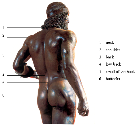
Figure 8: Human body, posterior view
TRUNK: SPINE, BACK, LOWBACK, CHEST, BREAST, RIBCAGE, ABDOMEN
The spine is the central support for the body. Another word for the spine is the backbone. The spine is made of separate irregular bones called vertebrae. There is a layer of cartilage (disc) in between each vertebra that keeps the bones from rubbing against each other. There are twenty six vertebrae in the spine. According to the region and position, the spine is divided into cervical – 7 vertebrae (C1-C7), thoracic – 12 vertebrae (T1-T12), lumbar – 5 vertebrae (L1-L5), sacral – 5 (fused) vertebrae (S1-S5) and coccygeal – 3-5 vertebrae (Co1-Co5).
The chest is the region of the body between the neck and the abdomen. The muscles covering the abdominal wall are abdominal muscles or abs. Ribs are the long curved bones, which form the rib cage. Ribs surround the chest (Latin thorax) and protect the lungs, heart, and other internal organs of the thoracic cavity. The muscles associated with this area are pectoral muscles or pecs and trapezius muscle.
LOWER LIMB: HIP, BUTTOCKS , LEG, THIGH, KNEE, CALF, LOWER LEG, ANKLE, FOOT, HEEL, TOES
The buttocks are formed by the masses of the gluteal muscles or glutes, superimposed by a layer of fat.
The leg is the lower extremity (limb) of the body, extending from the hip to the ankle, and including the thigh, the knee and the lower leg.
The thigh is the area between the hip and the knee. The single bone in the thigh is called the femur. This bone is very thick and strong and forms a ball and socket point at the hip, and a condylar joint at the knee.
The knee is the lower extremity joint connecting the femur and the tibia. It actually is comprised of two separate joints. The femoro-patellar joint consists of the patella, or "kneecap"and the patellar groove on the front of the femur through which it slides. The femoro-tibial joint links the femur, or thigh bone, with the tibia, the main bone of the lower leg. The cartilaginous elements within the knee joint, which serve to protect the ends of the bones from rubbing on each other, are called the menisci.
The lower leg is the area between the knee and the ankle, consisting of two bones, tibia and fibula. The muscles found at the back of the lower leg form the calf.
The ankle joint is formed where the foot and the leg meet. The ankle is a hinge joint that connects the distal ends of the tibia and fibula in the lower leg with the proximal end of the talus bone in the foot.
The foot, adjusted to bear the weight of the body, is made up by five toes (big, pointer, middle, ring, little or pinky toes), the bottom of the foot, that is called the sole, instep, the arch of the foot, and heel. The ball of the foot is where the toes join with the rest of the foot. It is muscular and easily blistered. Runners often move with their weight on the balls of their feet for better balance.
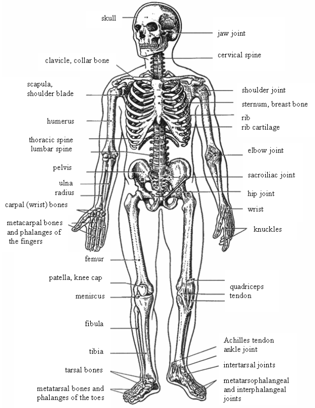
Figure 9: Skeletal system, anterior view
The human musculoskeletal system consists of the skeleton, made up of bones attached to other bones with joints, and skeletal muscles attached to the skeleton by tendons. The skeleton of an adult consists of more than 200 bones of various shapes and sizes. They are made up of hard osseous tissue and described as long, short, flat and irregular.
The human body contains more than 650 individual muscles which are attached to the skeleton by tendons. The main function of all muscles is to provide movement for the body. The muscular system consists of three different types of muscle tissues: skeletal, cardiac and smooth. Each of these different tissues has the ability to contract, which then allows body movements and functions. There are two types of muscles in the system and they are the involuntary muscles, and the voluntary muscles. The muscles working under our conscious control are called the voluntary muscles and the ones the function of which can not be consciously controlled are the involuntary muscles. The heart, or the cardiac muscle, is an example of an involuntary muscle.
SKELETAL
MUSCLES:
The skeletal muscles make up about 40 % of an adults body weight. It
has stripe-like markings, or striations. The skeletal muscles are
composed of long muscle fibers. The nervous system controls the
contraction of the muscle. Many of the skeletal muscle contractions are
automatic. However we still can control the action of the skeletal
muscle. And it is because of this reason that the skeletal muscle is
also called voluntary muscle.
SMOOTH
MUSCLES:
Most of our internal organs are made up of smooth muscles. They are
found in the urinary bladder, gallbladder, arteries, and veins. Also
the digestive tract is made up of smooth muscle as well. The smooth
muscles are controlled by the autonomic nervous system and hormones. We
cannot consciously control the smooth muscles and that is why they are
often called involuntary muscles.
CARDIAC
MUSCLE:
The cardiac muscle is a type of an involuntary striated muscle found
exclusively within the heart. Its function is to "pump" blood through
the circulatory system by contracting. Unlike skeletal muscle, which
contracts in response to nerve stimulation, cardiac muscle’s
function is based on self-excitable stimulating contraction without an
electrical impulse coming from the central nervous system.
Muscles generally work in pairs to produce movement: when one muscle flexes (or contracts) the other relaxes, a process known as antagonism.
An extensor muscle is any skeletal muscle that opens a joint increasing the angle between components of a limb, such as straightening the knee or elbow and bending the wrist or spine. With the exception of the knee joint the movement is directed backward. This action is known as extension.
A flexor muscle is a skeletal muscle whose contraction bends a joint, decreasing the angle between components of a limb, such as bending the knee or elbow. This action is known as flexion.
An abductor muscle is any of the muscles that cause movement of an extremity (limb) away from the midline of the body or away from a neighbouring part or limb.
An adductor muscle is any of the muscles that draw a part of the body toward its median line or toward the axis of an extremity.
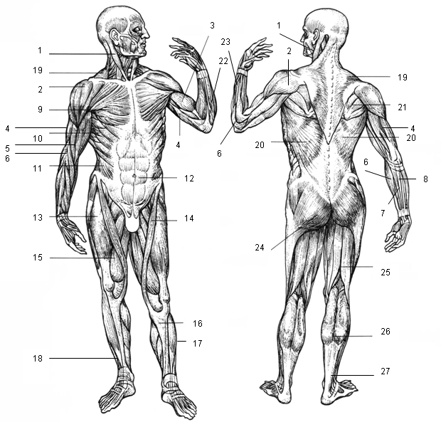
Figure 10: Muscular system, anterior and posterior view

Structurally, the nervous system is composed of two main parts:
- The central nervous system which consists of the brain and spinal cord.
- The peripheral nervous system, the spinal and cranial nerves.
There are also two functional subdivisions of the peripheral nervous system:
- somatic nervous system associated with the voluntary control of body movements through the action of skeletal muscles, and also reception of external stimuli.
- autonomic nervous system is the involuntary nervous system associated with regulation of the activities of visceral organs and glands. It has two divisions: sympathetic and parasympathetic system.
Neurons may be classified according to their function as:
- motor neurons (motoneurons); these neurons are efferent are carry motor impulses from the brain or spinal cord to the muscles, or organs to initiate activity,
- sensory neurons; these neurons are afferent and carry sensory impulses from a body part to the brain or spinal cord,
- connecting neurons, which transmit impulses from one part of the brain to another.
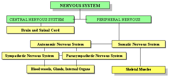
Figure 11: Organization of the Nervous System
In all skeletal muscles, contraction is stimulated by electrical impulses transmitted by the nerves, and motor neurons (efferent neurons, motoneurons) in particular. Neurons are basic nerve cells consisting of three parts: cell body, dendrite and axon. Dendrites are extensions from the cell body which conduct impulses to the cell body, axons are extensions carrying the impulses away from the cell body. Neurons communicate with one another via synapses. A synapse is a microscopic space between an axon and a dendrite. Chemicals which help an impulse cross the synapse (such as acetylcholine and catecholamine) are called neurotransmitters.
Motor neurons innervate or activate muscles groups to perform. A single motoneuron may synapse with one or more muscle fibers. One motoneuron and all of the muscle fibers to which it connects is a motor unit. Groups of motor units often work together to coordinate contractions of a single muscle; the number of muscle fibers within each unit can vary. Thigh muscles can have a thousand fibers in each unit, eye muscles might have only ten. In general, the number of muscle fibers innervated by a motor unit is a function of a muscle's need for refined motion. Muscles requiring more refined motion are innervated by motor units that synapse with fewer muscle fibers.

Figure 12 : Internal Organs
The term sports injury, in the broadest sense, refers to the kinds of injuries that most commonly occur during sports or exercise. Some sports injuries result from accidents, others are due to poor training practices, improper equipment, lack of conditioning, or insufficient warm-up and stretching.
Although any part of your body can be injured during sports or exercise, the term is usually reserved for injuries that involve the musculoskeletal system, which includes the muscles, bones, and associated tissues like cartilage.
A strain is an injury which occurs to a muscle in which the muscle fibers tear as a result of overstretching. Strains are also colloquially known as pulled muscles. The equivalent injury to a ligament is a sprain. Typical symptoms of a strain include localized pain, stiffness, swelling, inflammation, and bruising around the strained muscle.
Strains can happen to anyone and are certainly not restricted to athletes; nevertheless, people who are involved in sports are more at risk of developing a strain.
A sprain is an injury which occurs to ligaments caused by a sudden overstretching. The ligament is usually only stretched, but sometimes it can be snapped, slightly torn, or ruptured, all of which are more serious and require longer to heal.
Sprains are graded in three degrees. Although some signs and symptoms can be used to assess the severity of a sprain, the most definitive method is with the use of Magnetic Resonance Imaging (MRI). A first degree sprain has only minor tearing of the ligament whereas a third degree sprain is characterized by complete rupture.
The typical signs and symptoms associated with a sprain are the cardinal signs of inflammation: localized pain, swelling, and loss of function.
Although any joint can experience a sprain, some of the more common include the ankle, knee, and fingers. Perhaps one of the more spoken about sprains is that to the Anterior Cruciate Ligament of the knee. This is a disabling sprain common to athletes, especially basketball, soccer, and judo players.
Sprains can best be prevented by proper use of safety equipment (wrist, ankle guards), warm-ups and cool-downs (including stretching), being aware of your surroundings and maintaining strength and flexibility.
Achilles tendon injuries refer to a stretch, tear, or irritation to the tendon connecting the calf muscle to the back of the heel. The most common cause of Achilles tendon tears is a problem called tendinitis, a degenerative condition caused by aging or overuse. When a tendon is weakened, trauma can cause it to rupture.
A bone fracture is a medical condition in which a bone becomes cracked, splintered, or bisected as a result of physical trauma. In orthopaedic medicine, fractures are classified as closed or open (compound) and simple or multi-fragmentary (formerly comminuted).
Closed fractures are those in which the skin is intact, while open (compound) fractures involve wounds that communicate with the fracture and may expose bone to contamination. Open injuries carry an elevated risk of infection. They require antibiotics treatment and usually urgent surgical treatment. This involves removal of all dirt, contamination, and dead tissue.
Simple fractures are fractures that occur along one line, splitting the bone into two pieces, while multi-fragmentary fractures involve the bone splitting into multiple pieces. A simple, closed fracture is much easier to treat and has a much better prognosis than an open, contaminated fracture. Other considerations in fracture care are displacement and angulation. If angulation or displacement is large, reduction (manipulation) of the bone may be required and, in adults, frequently requires surgical care.
Stress fractures occur largely in the weight-bearing bones, such as the tibia or fibula (bones of the lower leg) and metatarsals (bones of the foot), and are common in sports that require repetitive impact, primarily running/jumping sports such as gymnastics or track and field. Running creates forces two to three times a person's body weight on the lower limbs.
Stress fractures usually have a narrow list of symptoms. It could present as a generalized area of pain, tenderness, and pain with weight-bearing. Usually when running, a stress fracture has severe pain in the beginning of the run, moderate pain in the middle of the run, and severe pain at the end and after the run. X-rays usually do not show any evidence of stress fractures, so a CT scan, or MRI may be more effective in unclear cases.
Joint dislocation takes place when bones in a joint become displaced or misaligned. It is often caused by a sudden impact to the joint. The ligaments almost always become damaged as a result of a dislocation. Once a joint is dislocated, it may reduce (return to its proper position) on its own, or it may require physical manipulation. Once reduction is achieved, the joint is held in place through a splint (for straight joints like fingers and toes) or a bandage (for complex joints like shoulders). Even if a dislocated joint reduces on its own, it should be immobilized and medical attention should be sought. Contact sports such as football and basketball, as well as high-impact sports and sports that can result in excessive stretching or falling, cause the majority of dislocations. The shoulders, fingers, and wrists are all common places for a dislocation to occur.
Menisci are cartilaginous elements within the knee joint which serve to protect the ends of the bones from rubbing on each other and to effectively deepen the tibial sockets into which the femur attaches. There are two menisci in each knee, the medial and the lateral meniscus. Either or both may be cracked, or torn, when the knee is forcefully rotated and/or bent.
Overtraining occurs when the volume and intensity of an exercise exceeds the organism’s recovery capacity. Improvements in strength and fitness occur only during the rest period following the training. This process takes at least 36 hours to complete. If sufficient rest is not available then complete regeneration cannot occur. If this imbalance between excess training and inadequate rest persists then the individual's performance will eventually plateau and decline. Overtraining may be accompanied by one or more of the following symptoms: persistent muscle soreness, persistent fatigue, elevated resting heart rate, increased susceptibility to infections, increased incidence of injuries, irritability, depression and loss of motivation.
Fortunately, most sports injuries can be treated effectively, and most people who suffer injuries can return to a satisfying level of physical activity after an injury. Even better, many sports injuries can be prevented if people take the proper precautions.
Reference:
The RICE Method
Questions:
- What does the acronym RICE mean?
- Why is the R-step that important as the first thing we have to do?
- What is the maximum period of time the I-step should be applied for? Why is there a limitation?
- What must be avoided when applying the C-step?
- What must be done to reach the appropriate E-step effect?
- What is the sign the RICE method may not be effective enough?
Achilles tendon Achillova šlacha
abdomen
břicho
abdominal
břišní
abdominals, abs břišní svaly
angulation
odklon od osy, zahnutí
ankle
kotník, hlezenní kloub
anus řiť
appendix
červovitý přívěsek slepého
střeva, apendix
arm paže
attach
upínat se, připojit
axon
výběžek (dlouhý) neuronu, neurit
back
záda
back muscles
zádové svalstvo
backbone
páteř
ball of the foot přední část
chodidla,
bříško
bend
ohýbat, ohnout, krčit, pokrčit
biceps biceps
bone kost
bronchus pl. bronchi průduška pl.
průdušky
bronchial stem bronchiální kmen
bruising
zhmoždění, krevní výron
buttocks
hýždě
caecum
slepé střevo
calf pl. calves lýtko
carpal bones zápěstní kosti
cartilage
chrupavka
cavity dutina
cervical
krční
chin brada
clavicle
klíční kost
coccygeal
kostrční
colon
tlusté střevo
ascending colon
vzestupný tračník
tlustého střeva
descending
colon sestupný tračník
tlustého střeva
sigmoid colon
esovitá klička tlustého střeva
transverse
colon příčný tračník
tlustého střeva
colloquial
hovorový, hovorový výraz
complementary space komplementární
prostor
conscious
vědomý
cool-down závěrečné
zklidnění,
vyklusání, protažení a
kompenzační cvičení
crack
prasknout, zlomit se
CT scan počítačová tomografie
dendrite
výběžek (krátký) neuronu,
dendrite
diaphragm
bránice
dislocation
dislokace, vykloubení
displace
posunout se (kosti při zlomenině), dislokovat
displacement
posun kostí při zlomenině, dislokace
distal
distální
duodenum
dvanáctník, část
tenkého střeva
esophagus
jícen
extend
natáhnout, napnout
extension
extenze, napnutí, propnutí,
natažení, výběžek (nervové buňky)
extensor
sval způsobující extenzi v kloubu
extremity
končetina
lower extremity
dolní končetina
upper extremity
horní končetina
fatigue
únava
femur kost
stehenní
fibula
lýtková kost
flex
pokrčit, ohnout
flexion
flexe, pokrčení, ohnutí
flexor sval
způsobující flexi v kloubu
forearm
předloktí
forehead čelo
fracture
fraktura, zlomenina
closed fracture
zavřená zlomenina
comminuted
fracture roztříštěná
zlomenina
compound
fracture otevřená, komplikovaná
zlomenina
displaced
fracture dislokovaná zlomenina
open fracture
otevřená zlomenina
simple fracture
jednoduchá, nekomplikovaná
zlomenina
stress fracture
únavová zlomenina
gallbladder
žlučník
gluteals, glutes hýžďové svaly
groin
tříslo
guards
chrániče
hairline fracture vlasová zlomenina,
naštípnutí
hamstrings hamstringy, svaly zadní strany stehna
handedness preference
užívání
pravé či levé končetiny, pravorukost, levorukost
left-handed levoruký,
levák
right-handed
pravoruký, pravák
heal hojit
(se), léčit (se)
heel pata
humerus kost
pažní
ileum
kyčelník, část tenkého střeva
imbalance
dysbalance
incidence
výskyt
inflammation
zánět
injury
zranění, úraz
innervate
inervovat
instep
nárt
intestine
střevo
large intestine
tlusté střevo
small intestine
tenké střevo
irritability
podrážděnost
jejunum
lačník, část tenkého střeva
joint kloub
knee koleno
knuckle
kloub prstu
larynx hrtan
ligament vaz
limb
končetina
liver
játra
lower leg
bérec
lumbar
bederní
lungs
plíce
meniscus pl. menisci meniskus
mesenterium
mezenterium, okruží
metacarpal bones záprstní kosti
metatarsal bones zánártní
kosti
misalign
být v nesprávném
postavení
motor unit motorická jednotka
MRI (Magnetic Resonance Imaging) magnetická
rezonance
multi-fragmentary mnohočetný
muscle sval
adductor
magnus muscle [ә׀daktә] velký přitahovač stehna
anterior
tibial muscle [æn׀tiәriә tibiәl]
přední
sval holenní
biceps
brachii, biceps of the arm [baisәps breikiai]
dvojhlavý sval pažní
biceps of the
thigh dvojhlavý sval stehenní
brachial muscle
[breikiәl] ohýbač paže
brachioradial
muscle [breikiә׀reidiәl] sval vřetenní
com. extensor
of fingers natahovač prstů
deltoid muscle
deltový sval
deltoid muscle
sval deltový
external
oblique abdominal muscle [ә׀bli:k] zevní
šikmý sval břišní
flexor of
fingers ohýbač prstů ruky
flexor of the
wrist ohýbač ruky
gastrocnemius
[gæstrә׀kni:miәs] sval
lýtkový
gluteus
maximus, greatest gluteal muscle velký sval
hýžďový
greater
pectoral muscle, pectoralis major velký
prsní sval
infraspinous
muscle [infrә׀spainәs] podhřebenový sval
involuntary
muscle hladký sval
latissimus
dorsi [lætisimәs do:sai], broadest of the back
široký sval zádový
long extensor
of toes dlouhý natahovač prstů
long peroneal
[perә׀ni:әl]muscle dlouhý sval
lýtkový
quadriceps
muscle čtyřhlavý sval stehenní
rectus
(straight) abdominal muscle přímý
břišní sval
sartorius
muscle [særtoriәs] sval krejčovský
serratus
anter. muscle [se׀reitәs] pilovitý sval
skeletal muscle
kosterní, příčně
pruhovaný sval
sternocleidomastoid [stә:nәklaidә׀mæstoid]
zdvihač hlavy
trapezius
muscle [trә׀pi:ziәs] sval trapézový
triceps of the
arm [traiseps] trojhlavý sval pažní
triceps of the
arm trojhlavý sval pažní
ulnar extensor
of the wrist [alnә] natahovač ruky
voluntary
muscle kosterní, příčně
pruhovaný sval
musculoskeletal
muskuloskeletální,
pohybový
nervous system nervový systém
autonomic
nervous system autonomní nervový
systém
neuron
neuron, nervová buňka
afferent neuron
aferentní, vzestupný,
dostředivý neuron
efferent neuron
eferentní, sestupní,
odstředivý neuron
motor neuron
motorický, hybný neuron, motoneuron
sensory neuron
sensitivní neuron
neurotransmitter
neurotransmiter
occur
objevovat se, vyskytovat se
overstretching
nadměrné protažení,
natažení
overtraining
přetrénování
overuse
přetížení,
přetěžování
pain bolest
severe pain
velká, krutá bolest
moderate pain
středně silná, snesitelná bolest
pancreas
slinivka břišní
patella
čéška
pectoral
prsní
pectoral
muscles prsní svaly
pectorals, pecs
prsní svaly
pelvis
pánev
persistent
přetrvávající,
dlouhodobý
phalanges
kosti prstů
pharynx hltan
plateau
plateau, stagnace
pleura
poplicnice
pleural cavity pleurální
štěrbina
proximal
proximální
quadriceps
kvadriceps, čtyřhlavý sval stehenní
radius
vřetenní kost
reduce
omezit, zmenšit
reduction
zmenšení
rib žebro
rib
cage
hrudní koš
rupture
ruptura, přetržení
sacral
křížový
sacroiliac joint křížokyčelní
skloubení
scapula
lopatka
severing
závažné poškození
severity
závažnost
skeleton
kostra
skull lebka
sliver
naštípnout, štěpit
small of the back kříž
snap
prasknout
sole chodidlo
soreness
bolest, bolestivost
spinal
páteřní,
týkající se páteře
spinal cord páteřní
mícha
spine
páteř
cervical spine
krční páteř
coccygeal
kostrč, kostrční obratle
lumbar spine bederní
páteř
sacral spine
křížová páteř
thoracic spine
hrudní páteř
spleen
slezina
splint dlaha
sprain
výron, distorze
sternum
prsní kost
stiffness
ztuhlost
strain
natržení
susceptibility
náchylnost, tendence k něčemu
swelling otok
tarsal bones nártní kosti
tear
natrhnout, přetrhnout
tendon
šlacha
thigh stehno
thoracic
hrudní
thumb palec
ruky
tibia
holenní kost
toe prst nohy
trachea
průdušnice
tracheal hilum
[hailәm] pl.
hila
plicní hilus
treatment
léčba
triceps
triceps
ulna
loketní kost
vertebra pl.
vertebrae
obratel, pl.
obratle
volume objem
warm-up rozcvičení,
rozehřátí
weight-bearing nosný (kost, kloub)
wrist
zápěstí
X-rays roentgen